Meeting
Place & Times:
EEF 2405
Wednesdays 13.00-16.00
Office
Hours:
EEF 7205 only
by
appointment either
by e-mail to cilesiz[at]itu.edu.tr or
by phone to 212 285 67 73
|
BYM504E Biomedical
Imaging Systems (CRN: 23514)
Grading: Simulation/Assignments 20%;
Midterm Exam 20%; Project 20%; Final Exam 40%
|
Midterm Exam:
|
tentatively 9th week of
classes |
|
Imaging Simulation due:
|
tentatively after the
BREAK (26-30 March 2018)
|
|
Project Presentations:
|
9
and 16 May 2018
|
|
Final Exam:
|
TBA
|
There IS a
NINOVA page for this course! |
|
Course
Objective:
|
This course will provide a
detailed review of imaging principles and instrumentation of all the
conventional clinical imaging systems, including X-ray radiography,
computerized tomography (CT), gamma camera, SPECT, PET, ultrasound (US),
Doppler US, Magnetic Resonance (MR) and functional MR (f-MR).
|
|
Course
Description:
|
• General characteristics of
imaging systems;
• X-ray and CT: general principles,
interaction of X-rays with tissues, contrast agents, imaging techniques,
image reconstruction, radiation dose;
• Nuclear Medicine: general
principles, radionuclide, radioactive decay, gamma camera, imaging
techniques, SPECT, PET;
• Ultrasound imaging: general
principles, interaction of acoustic waves with tissue, acoustic impedance,
instrumentation, scanning modes, artifacts, blood velocity measurements,
contrast agents;
• MR imaging: general principles,
nuclear magnetism, magnetic resonance, instrumentation, imaging sequences,
contrast agents, imaging techniques, functional MRI.
|
|
Participation/Assignments:
|
• As part of in class
participation each student will have to do several oral and/or written
presentations on topics assigned by the instructor.
• Each student will simulate an imaging modality (such as, CT or US) using a
tissue phantom of his/her choice and will present the results with a
demonstration and in laboratory report format.
• An in depth review on an imaging modality/system not covered in class.
Results of this literature/lab or clinical study review will be presented
orally in class and in writing in report format.
|
|
Course Plan:
|
|
Weeks |
Topics |
|
1 |
Introduction to
biomedical imaging |
|
2 |
General image characteristics |
|
3 |
X-rays, X-ray
film, instrumentation |
|
4 |
CT,
instrumentation, Fourier slice theorem, image reconstruction |
|
5 |
Nuclear
medicine, radioactivity, technetium generator, use of technetium |
|
6 |
Gamma camera,
SPECT, PET, instrumentation |
|
7 |
Image
reconstruction, clinical applications |
|
8 |
Ultrasound, wave
propagation and acoustic impedance, instrumentation |
|
9 |
Midterm exam |
|
10 |
US imaging
characteristics, scanning methods and modes, Doppler US |
|
11 |
MR imaging,
magnetic resonance, Larmor frequency, relaxation
|
|
12 |
Slice selection,
phase/frequency encoding, imaging sequences, functional MRI |
|
13 |
Project
presentations |
|
14 |
Project
presentations |
|
|
Textbooks:

|
Introduction to
Medical Imaging: Physics, Engineering and Clinical Applications,
Nadine B. Smith & Andrew Webb, Cambridge University Press, 2011, ISBN-13:
978-0521190657.
(shown on the left!)
Other
reference books:
Biomedical Imaging: Principles and Applications, Editor: Reiner
Salzer, John Wiley & Sons, Inc., 2012 , Online ISBN: 9781118271933.
The Chemistry of Molecular Imaging, Editors: Nicholas Long, Wing-Tak
Wong,
John Wiley & Sons, Inc., 2015, Online ISBN: 9781118854754.
Advances in Optical Imaging for Clinical Medicine, Editor(s):
Nicusor Iftimia, William R. Brugge, Daniel X. Hammer, John Wiley & Sons,
Inc., 2011, Online ISBN: 9780470767061.
Do
not FORGET to check out this link.
You need to have online access to library resources to read this handbook.
That also means you need to check out online library resources such as
Knovel E-Kitap
and
CRC ENGnetBASE.
|
|
Project:
|
involves an in depth review
on one of the newer biomedical imaging modalities not covered in class...
if available: principles,
instrumentation on the market, along with price and maintenance
are to be covered in a 15-20 minute in-class oral presentation on the last
two weeks of class AND an accompanying 10 page maximum word-processed report.
Reports without (i)
identification of its author, (ii) a reference list, and (iii) numerous
spelling errors (please run spell-check) lead to "reduced"
grades. Mot-a-mot copied (i.e., copy/paste) reports are not
favored! For information on how to write an effective report and make an
effective presentation consult "Scientific
and Technical Writing" lectures notes.
|
|
Suggested
Topics:
|
- new frontiers in Optical
Coherence Tomography
- new frontiers in
opto-acoustic/deep tissue Imaging
-
3-d Optical Projection
Tomography
-
Optical Metabolic
Imaging
-
T-ray Imaging
-
Magnetic Field
Correlation MR Imaging
-
DNP-enhanced MRI (DNP =
Dynamic Nuclear Polarization)
-
Near-infrared fluorescence
imaging (with or without biomarkers)
-
Endoscopic multispectral
imaging
-
Hybrid imaging
-
Dual imaging
-
Fusion imaging
ANYTHING ELSE NOT COVERED IN CLASS, but INTERESTING TO
YOU.
Other
topics may be found in the online books under CRC ENGNETBASE and
WILEY e-BOOKS available
from ITU library on and off-site using your
ID.
|
|
Interesting
Links:
|
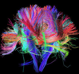
Human
Connectome Project (see and click on the images on the left!)
Micro CT
Dental MicroCT
Overview of Imaging Tests
Modern
Imaging Techniques
Physics and
Technology of Medical Imaging
Introduction to Ultrasonic Testing
Medical Imaging News
Medical Imaging Modalities
|
|
|
For more
info on class visit previous years' web sites in the archives...
|
|
|
|